Scaphoid fracture and avascular necrosis
The shot
In essence
Scaphoid fractures are the most common carpal injury and risk proximal pole avascular necrosis due to retrograde arterial blood supply. Untreated cases progress to non-union, dorsal intercalated segment instability, and scaphoid non-union advanced collapse.
Say this in the viva
I am presented with posteroanterior and lateral radiographs of the right wrist, there is an ununited transverse fracture through the proximal third of the scaphoid waist with approximately two millimetres of cortical displacement. The proximal pole demonstrates marked sclerosis and subtle flattening compared to the surrounding carpus, highly suspicious for avascular necrosis. On the lateral view, the scapholunate angle is increased to approximately 70 degrees with dorsal tilt of the lunate, consistent with a DISI instability pattern. There is no significant radioscaphoid joint space loss or osteophytosis to suggest established SNAC changes. On the subsequent coronal T1 and fat-suppressed T2-weighted MRI sequences, the proximal pole exhibits diffuse low T1 and low T2 signal, with complete absence of enhancement following intravenous gadolinium administration, confirming non-viable bone and avascular necrosis. The diagnosis is a proximal scaphoid non-union complicated by avascular necrosis and carpal instability. I would compare with any previous imaging, if available, and discuss this urgently with the orthopaedic hand multidisciplinary team for further assessment.
The money findings
- Retrograde arterial supply enters the dorsal ridge, placing proximal pole fractures at the highest risk of AVN (~100% in proximal fifth).
- Plain film features of AVN: relative or absolute sclerosis, cortical collapse, fragmentation, and cystic change in the proximal pole.
- Contrast-enhanced MRI is the definitive imaging modality to assess proximal pole vascularity and viability prior to surgery.
- Carpal instability: assess for DISI pattern (scapholunate angle >60 degrees, capitolunate angle >30 degrees with dorsal tilt of the lunate).
- SNAC wrist staging: Stage 1 (radial styloid), Stage 2 (scaphocapitate joint), Stage 3 (capitolunate joint); the radiolunate joint is characteristically spared.
- Fine-slice CT reconstructed along the long axis of the scaphoid is the gold standard for assessing union, displacement, and humpback deformity.
- Management: urgent referral to the orthopaedic hand multidisciplinary team.
The discriminators
- Preiser disease - idiopathic scaphoid osteonecrosis occurring without an antecedent fracture line.
- Transient relative osteosclerosis - apparent proximal pole sclerosis due to disuse osteopenia of the surrounding carpus, showing normal post-contrast MRI enhancement.
- Ununited scaphoid fracture without AVN - corticated fracture gap with preserved T1 marrow signal and robust post-contrast enhancement of the proximal pole.
Classification
SNAC wrist staging: Stage 1: radial styloid arthrosis. Stage 2: scaphocapitate arthrosis. Stage 3: capitolunate joint arthrosis (radiolunate joint characteristically spared).
Management
Urgent referral to the specialist orthopaedic hand multidisciplinary team; advise fine-slice wrist CT with scaphoid long-axis reconstructions to quantify deformity and bridging, alongside contrast-enhanced MRI to determine proximal pole vascular viability.
Clinical buzz words
The trap
Mistaking transient relative osteosclerosis caused by adjacent disuse osteopenia for true osteonecrosis, which requires contrast-enhanced MRI rather than non-contrast imaging to definitively prove non-viability.
Check yourself
1. On MRI, what findings confirm proximal pole avascular necrosis and non-viable bone in scaphoid fracture?
Show the answerHide the answer
2. How is Preiser disease distinguished from scaphoid fracture non-union complicated by avascular necrosis?
Show the answerHide the answer
3. Describe the three stages of scaphoid non-union advanced collapse wrist arthrosis.
Show the answerHide the answer
4. What is the diagnostic pitfall when evaluating apparent scaphoid proximal pole sclerosis?
Show the answerHide the answer
Cases on Radiopaedia
 Scaphoid avascular necrosisRadiopaedia
 Avascular necrosis - scaphoid non-unionRadiopaedia
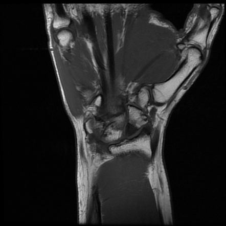 Avascular necrosis of scaphoidRadiopaedia
 Scaphoid avascular necrosis (contrast-enhanced MRI)Radiopaedia
 Normal wrist alignment, dorsal and volar intercalated segmental instability (illustration)Radiopaedia
More cases of scaphoid fracture and avascular necrosis on Radiopaedia
Sign in to keep track of the lessons you have done.