Lisfranc injury

The shot
In essence
Disruption of the tarsometatarsal (TMT) joint complex ranging from subtle ligamentous injury to gross fracture-dislocation. Missed injuries lead to chronic midfoot instability, longitudinal arch collapse, and debilitating secondary osteoarthritis.
Say this in the viva
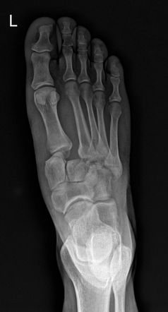
I am presented with weight-bearing dorsoplantar and oblique radiographs of the right foot in an adult. On the dorsoplantar view, there is widening of the space between the first and second metatarsal bases, measuring approximately four millimetres. Crucially, there is lateral displacement of the second metatarsal base, which no longer aligns with the medial border of the intermediate cuneiform. In the first intermetatarsal space, there is a small, well-corticated avulsion bone fragment representing a classic fleck sign. On the oblique projection, there is also mild lateral step-off at the third tarsometatarsal joint. The lateral view demonstrates preserved overall alignment without gross dorsal dislocation or flattening of the longitudinal arch, and the visualized ankle joint and calcaneus are intact. These findings represent an unstable Lisfranc tarsometatarsal fracture-dislocation. I would compare with any previous imaging, if available, recommend the patient remains non-weight-bearing, arrange an urgent CT scan of the midfoot to fully characterize the fracture anatomy and occult subluxations, and urgently refer to the on-call orthopaedic team.
The money findings
- Widening >2 mm between the bases of the 1st and 2nd metatarsals on dorsoplantar radiographs is abnormal.
- Medial border of the 2nd metatarsal base must align with the medial border of the intermediate (middle) cuneiform on the AP view.
- Medial border of the 3rd metatarsal base must align with the medial border of the lateral cuneiform on the 30-degree oblique view.
- Fleck sign: tiny avulsion bone fragment in the 1st-2nd intermetatarsal space pathognomonic of Lisfranc ligament avulsion.
- The Lisfranc ligament attaches the plantar-lateral medial cuneiform to the plantar-medial base of the 2nd metatarsal.
- Myerson classification: Type A (homolateral/total incongruity), Type B (partial incongruity), Type C (divergent).
- Non-weight-bearing radiographs miss up to 50% of subtle injuries; weight-bearing views, CT, or MRI are required.
- Next step: keep patient non-weight-bearing, arrange CT midfoot, and urgently refer to orthopaedics.
The discriminators
- Isolated metatarsal base fracture - metatarsal fracture line is present but normal alignment of the 2nd and 3rd TMT lines is strictly preserved
- Charcot neuroarthropathy - marked subchondral sclerosis, joint fragmentation, and rocker-bottom deformity in a neuropathic foot
- Low-grade midfoot sprain - midfoot pain following trauma but normal TMT alignment maintained with no diastasis >2 mm or fleck fragment
Classification
Myerson classification: Type A (homolateral/total incongruity), Type B (partial incongruity), Type C (divergent)
Management
Advise non-weight-bearing status, arrange urgent CT midfoot for detailed assessment, and refer urgently to the on-call orthopaedic team.
Clinical buzz words
The trap
Dismissing the tiny fleck sign in the 1st intermetatarsal space as an incidental ossicle, missing an unstable ligamentous injury.
Check yourself
1. What normal anatomical alignment must be maintained at the second tarsometatarsal joint on a dorsoplantar foot radiograph?
Show the answerHide the answer
2. How is an isolated metatarsal base fracture distinguished from a Lisfranc injury?
Show the answerHide the answer
3. What categories comprise the Myerson classification for Lisfranc injuries?
Show the answerHide the answer
4. What is the critical diagnostic trap when reviewing radiographs for a suspected Lisfranc injury?
Show the answerHide the answer
Cases on Radiopaedia
 Lisfranc injuryRadiopaedia
 Lisfranc injuryRadiopaedia
 Lisfranc injuryRadiopaedia
 Lisfranc injury - Myerson classifications (illustrations)Radiopaedia
More cases of lisfranc injury on Radiopaedia
Sign in to keep track of the lessons you have done.