Pulmonary sequestration

The shot
In essence
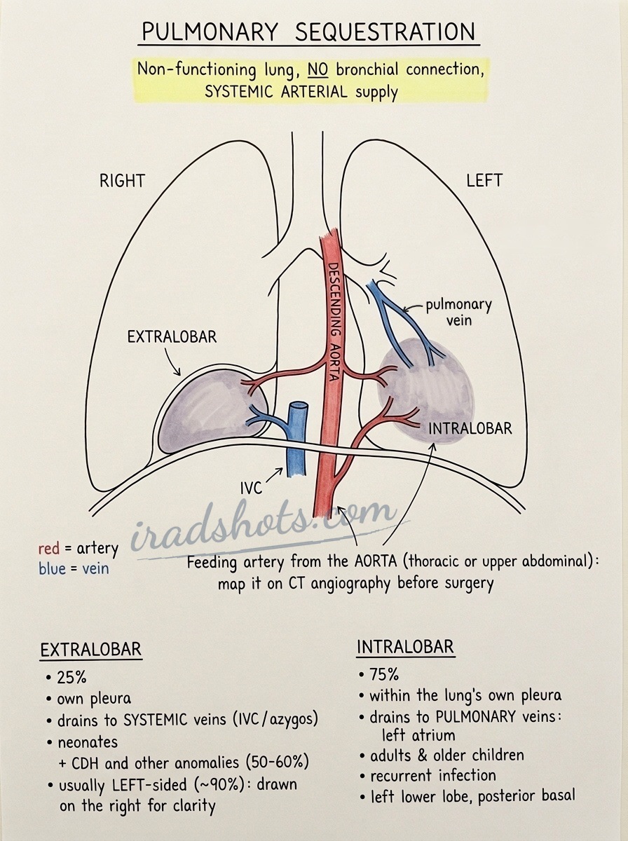
Pulmonary sequestration is a congenital bronchopulmonary foregut malformation comprising non-functioning dysplastic lung tissue that lacks communication with the tracheobronchial tree and receives systemic arterial supply, most commonly from the descending aorta.
Say this in the viva
I am presented with a chest radiograph that demonstrates a well-defined opacity in the left lower zone situated within the retrocardiac space and medial posterior costophrenic sulcus. On the subsequent contrast-enhanced CT of the chest, there is a well-circumscribed, multicystic soft-tissue mass in the posterior basal segment of the left lower lobe. Crucially, I can identify a large anomalous systemic feeding artery arising directly from the descending thoracic aorta to supply the lesion, with venous drainage directed into the inferior pulmonary veins. There is no communication with the normal tracheobronchial tree, and there is no evidence of chest wall invasion, destructive osseous change, or suspicious lymphadenopathy. These findings are diagnostic of an intralobar pulmonary sequestration. A congenital pulmonary airway malformation or a persistent lower lobe abscess would not demonstrate an anomalous systemic arterial supply unless a hybrid lesion is present. I would compare with any previous imaging, if available, and my next step is to recommend discussion at the thoracic multidisciplinary team meeting, explicitly highlighting the precise anatomical origin and course of the systemic feeding vessel on CT angiography to aid preoperative vascular road-mapping and mitigate the risk of severe intraoperative haemorrhage.
The money findings
- Location: medial/posterior basal segment of the left lower lobe is the classic site (~60%).
- Defining hallmark: anomalous systemic arterial supply, usually from descending thoracic or upper abdominal aorta.
- Intralobar (75%): shares normal visceral pleura, drains to pulmonary veins, presents in older children/adults with recurrent infection.
- Extralobar (25%): separate visceral pleura, drains to systemic veins (azygos/IVC), presents in neonates, 50% have associated anomalies (e.g. CDH).
- Hybrid lesion: features of CPAM (cystic architecture) combined with a systemic feeding vessel.
- Crucial risk: catastrophic haemorrhage if the systemic feeding artery is unrecognized.
The discriminators
- Congenital pulmonary airway malformation (CPAM) - lacks an anomalous systemic arterial supply and is supplied by the normal pulmonary circulation unless a hybrid lesion.
- Persistent pneumonia / lung abscess - supplied by normal pulmonary arterial vasculature rather than an anomalous systemic branch from the aorta.
- Congenital diaphragmatic hernia - visible diaphragmatic defect with herniation of abdominal bowel loops or omental fat into the hemithorax.
Classification
Intralobar (75%): shared visceral pleura, pulmonary venous drainage, presents in older children/adults with infection.
Extralobar (25%): separate visceral pleura, systemic venous drainage (azygos/IVC), presents in neonates, 50% associated anomalies (e.g. CDH).
Management
Discuss at thoracic or paediatric MDT. Perform multiphase CT angiography (including the upper abdomen) for definitive vascular road-mapping to demonstrate arterial supply and venous drainage.
Clinical buzz words
The trap
Failing to identify the anomalous systemic arterial feeder (which can arise below the diaphragm from the upper abdominal aorta) leading to fatal intraoperative haemorrhage.
Check yourself
1. What is the defining imaging hallmark of a pulmonary sequestration?
Show the answerHide the answer
2. How is a pulmonary sequestration differentiated from a congenital pulmonary airway malformation on cross-sectional imaging?
Show the answerHide the answer
3. How do intralobar and extralobar pulmonary sequestrations differ regarding their pleural coverage and venous drainage?
Show the answerHide the answer
4. What critical trap must be avoided when evaluating the arterial supply of a pulmonary sequestration?
Show the answerHide the answer
Cases on Radiopaedia
Find cases of pulmonary sequestration on RadiopaediaSketch
Sign in to keep track of the lessons you have done.