Pulmonary sarcoidosis
The shot
In essence
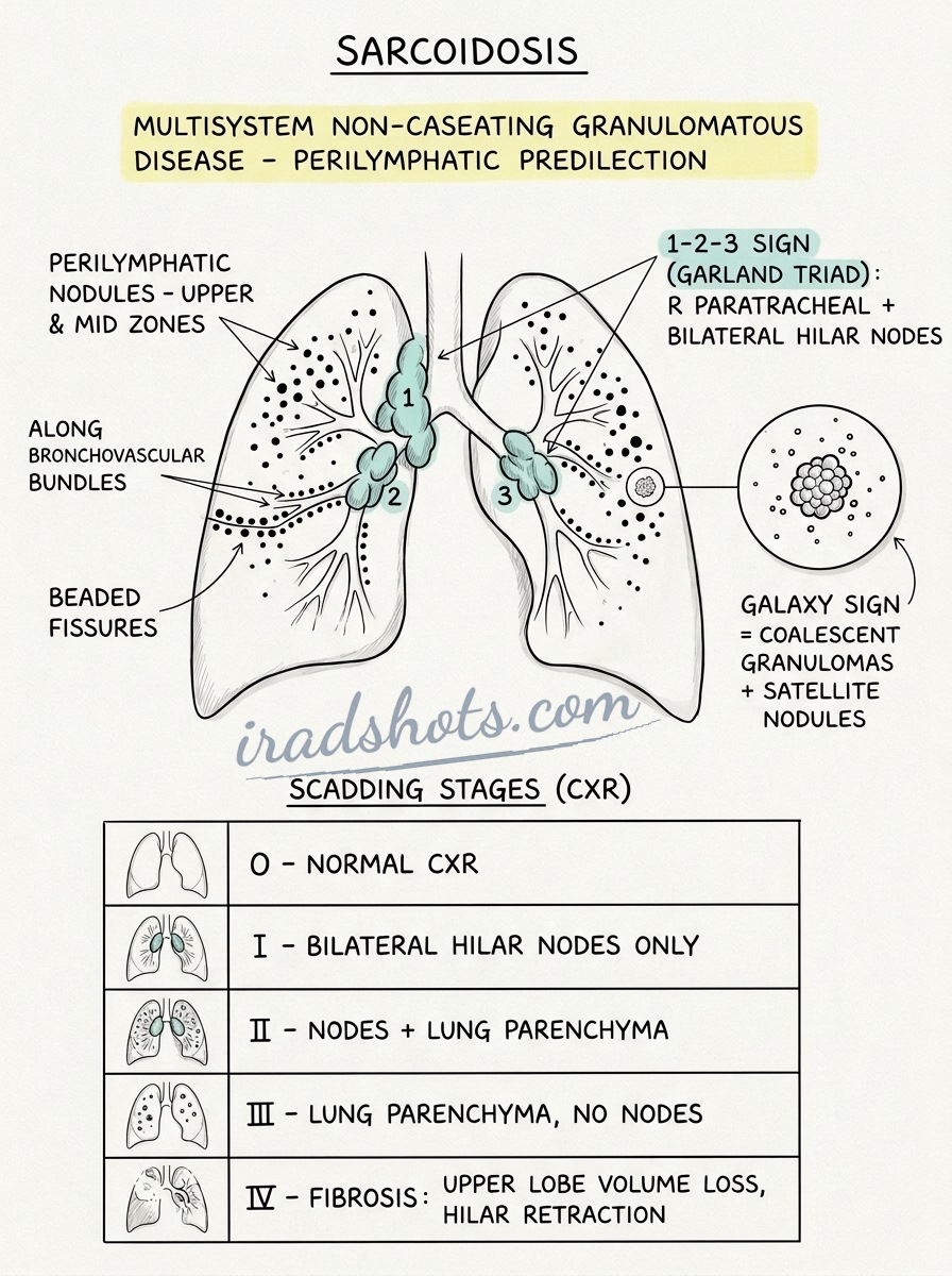
Pulmonary sarcoidosis is a multisystem non-caseating granulomatous disease with a predilection for the thoracic lymph nodes and lung parenchyma. It classically manifests as symmetric bilateral hilar and right paratracheal lymphadenopathy with upper/mid-zone perilymphatic nodularity.
Say this in the viva
I am presented with a chest radiograph demonstrating symmetrical bilateral hilar lymphadenopathy and right paratracheal widening, representing the classic 1-2-3 sign, alongside fine reticulonodular opacities predominantly in the mid- and upper lung zones. There is no pleural effusion or pneumothorax, and the lung bases are clear. On the accompanying HRCT chest, there is well-defined perilymphatic nodularity distributed along the bronchovascular bundles, subpleural surfaces, and major fissures, with upper- and mid-zone predominance. Symmetrical mediastinal and hilar lymphadenopathy is confirmed without low-attenuation central necrosis, and there is no evidence of architectural distortion, traction bronchiectasis, or honeycombing. These findings are characteristic of active pulmonary sarcoidosis, Scadding Stage II. The main differentials to consider are tuberculosis, though tuberculous lymphadenopathy is usually asymmetrical and hypoattenuating with centrilobular tree-in-bud opacities, and lymphoma, which lacks this characteristic perilymphatic nodular pattern. I would compare with any previous imaging, if available, to assess chronicity. I would suggest checking serum ACE and serum and urinary calcium levels to screen for hypercalcaemia, arranging pulmonary function tests, and performing a baseline ECG. Finally, I would refer the patient to the interstitial lung disease MDT, proposing EBUS-TBNA of the mediastinal nodes if histological confirmation is required.
The money findings
- Garland triad (1-2-3 sign): Symmetrical bilateral hilar and right paratracheal lymphadenopathy.
- Perilymphatic nodule distribution: Upper and mid-zone predilection along bronchovascular bundles, subpleural spaces, and fissures.
- Galaxy sign: Large central coalescent nodule surrounded by tiny satellite nodules.
- Scadding staging: 0 (normal), I (BHL only), II (BHL + parenchymal disease), III (parenchymal disease only), IV (end-stage fibrosis).
- Stage IV complications: Upper-lobe fibrocystic destruction predisposes to secondary aspergilloma (mycetoma) formation.
- Next step: Compare with prior imaging, assess calcium metabolism (ACE, serum/urinary calcium), and arrange EBUS-TBNA if histological proof is required.
The discriminators
- Tuberculosis - asymmetrical, hypoattenuating/necrotic lymphadenopathy with centrilobular tree-in-bud nodularity and cavitation
- Lymphoma - bulky anterior mediastinal nodal enlargement without characteristic perilymphatic nodularity
- Silicosis / Coal Workers' Pneumoconiosis - upper-zone nodules accompanied by a documented occupational exposure history
Classification
Scadding staging: Stage 0 (normal), Stage I (BHL only), Stage II (BHL + parenchymal disease), Stage III (parenchymal disease only), Stage IV (end-stage fibrosis)
Management
Check serum calcium and ACE levels, arrange pulmonary function tests with DLCO, and refer to respiratory MDT for EBUS-TBNA if histological proof is required.
Clinical buzz words
The trap
Calling unilateral hilar enlargement sarcoidosis; unilateral adenopathy is extremely rare in sarcoid and must be treated as malignancy or TB until proven otherwise.
Check yourself
1. What constitutes the Garland triad, or 1-2-3 sign, in pulmonary sarcoidosis?
Show the answerHide the answer
2. How can pulmonary sarcoidosis be distinguished from tuberculosis on thoracic imaging?
Show the answerHide the answer
3. Outline the Scadding staging system for pulmonary sarcoidosis.
Show the answerHide the answer
4. What is the key diagnostic trap when evaluating unilateral hilar lymphadenopathy?
Show the answerHide the answer
Cases on Radiopaedia
Find cases of pulmonary sarcoidosis on RadiopaediaSketch
Sign in to keep track of the lessons you have done.