Acute ischaemic stroke
The shot
In essence
Acute ischaemic stroke requires rapid identification of early parenchymal and vascular signs on unenhanced CT to exclude haemorrhage and assess eligibility for reperfusion therapies. Imaging determines vascular territory, ASPECTS score, and vessel occlusion status to guide hyperacute intravenous thrombolysis and mechanical thrombectomy.
Say this in the viva
I am presented with an unenhanced axial CT head of an adult patient. There is no evidence of acute intra- or extra-axial haemorrhage. In the right middle cerebral artery territory, there is loss of the right insular ribbon, subtle blurring of the right lentiform nucleus, and effacement of adjacent cortical sulci over the right frontotemporal convexity. A hyperdense right M1 segment MCA sign is evident. The remaining cortical grey-white matter differentiation and posterior fossa structures are preserved, giving an ASPECTS score of 8. These findings represent an acute right MCA territory ischaemic stroke secondary to an acute M1 segment thromboembolism. I would compare with any previous imaging, if available, and immediately telephone the acute stroke team to expedite hyperacute assessment. I would recommend an urgent CT angiogram from the aortic arch to the vertex to confirm the occlusion site and assess intracranial collateral circulation to determine suitability for emergency mechanical thrombectomy.
The money findings
- Unenhanced CT primary goal: exclude intracranial haemorrhage before reperfusion.
- Hyperdense vessel sign (e.g. hyperdense MCA or MCA dot sign) indicates acute intraluminal thrombus.
- Early parenchymal signs: loss of insular ribbon, lentiform blurring, cortical sulcal effacement, and loss of grey-white matter differentiation.
- ASPECTS score quantifies MCA territory ischaemia across 10 regions (3 subcortical, 7 cortical); score <6 suggests large core.
- CT angiogram from arch to vertex is required to confirm large vessel occlusion (LVO) and assess collateral status.
- CTA / CTP / MRI mismatch evaluates potential for mechanical thrombectomy (e.g. anterior circulation LVO within 6 hours, or up to 24 hours with imaging mismatch per NICE NG128).
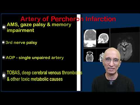
- Bilateral medial thalamic infarcts indicate occlusion of the variant Artery of Percheron.
The discriminators
- Vascular calcification - bilateral, discontinuous, and hyperdense (>100 HU) without parenchymal ischaemic changes.
- Post-ictal state (Todd paresis) - unenhanced CT is typically normal without territorial hypodensity or loss of grey-white differentiation.
- High-grade glioma - finger-like vasogenic oedema predominantly sparing the cortex with mass effect disproportionate to hyperacute onset.
Classification
ASPECTS (10 points total; deduct 1 per region involved): Ganglionic level (Caudate, Lentiform, Internal capsule, Insular ribbon, M1, M2, M3); Supraganglionic level (M4, M5, M6). Score <6 indicates large ischaemic core.
Management
Immediate direct telephone discussion with the hyperacute stroke team; recommend urgent CTA from aortic arch to vertex to assess large vessel occlusion and collateral score, and consider CT perfusion or MRI for wake-up or extended window (6-24 hour) presentations.
Clinical buzz words
The trap
Mistaking dense atheromatous wall calcification for a hyperdense MCA sign; verify whether attenuation exceeds 100 HU and check for bilateral symmetry.
Check yourself
1. In acute ischaemic stroke, what does a hyperdense vessel sign on unenhanced CT indicate?
Show the answerHide the answer
2. How can acute ischaemic stroke be distinguished from a high-grade glioma on CT?
Show the answerHide the answer
3. In the ASPECTS scoring system for acute middle cerebral artery ischaemic stroke, what threshold indicates a large core?
Show the answerHide the answer
4. What imaging trap can occur when evaluating for a hyperdense MCA sign in acute ischaemic stroke?
Show the answerHide the answer
Cases on Radiopaedia
 Acute ischemic stroke: dense MCA signRadiopaedia
 Wake-up ischemic strokeRadiopaedia
 Basilar tip thromboembolismRadiopaedia
 Loss of insular ribbon - sign of early MCA infarctionRadiopaedia
 Disappearing basal ganglia signRadiopaedia
 Carotid dissection with watershed infarctRadiopaedia
 Artery of Percheron InfarctionYouTube
More cases of acute ischaemic stroke on Radiopaedia
Sign in to keep track of the lessons you have done.