Cerebral venous sinus thrombosis
The shot
In essence
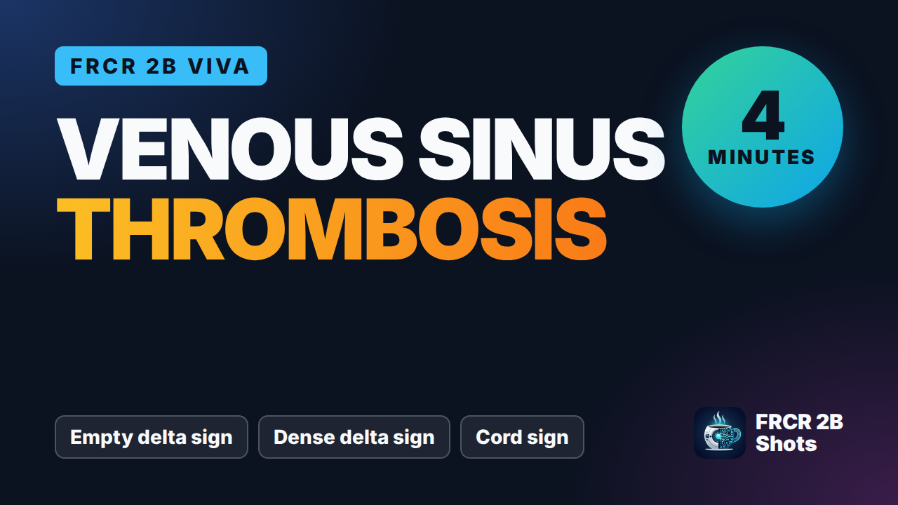
Cerebral venous sinus thrombosis (CVST) is a neurovascular emergency causing venous hypertension, vasogenic/cytotoxic oedema, and non-arterial territory haemorrhagic infarction. Imaging demonstrates hyperdense sinuses or cortical veins on unenhanced CT, filling defects ('empty delta sign') on CT or MR venography, and loss of normal flow voids on MRI.
Say this in the viva
I am presented with an axial non-contrast CT brain of a young adult female. There is abnormal low attenuation with loss of grey-white matter differentiation within the parasagittal high frontal cortex bilaterally, accompanied by small foci of petechial haemorrhage. These changes do not follow an arterial vascular territory. Additionally, there is hyperdensity within the posterior aspect of the superior sagittal sinus, representing the dense delta sign. On the subsequent post-contrast CT venogram images provided, there is a distinct non-enhancing filling defect within the superior sagittal sinus and the straight sinus, confirming the empty delta sign. There is no evidence of tonsillar herniation or midline shift. I would compare with any previous imaging, if available, but the findings are diagnostic of acute cerebral venous sinus thrombosis with secondary bilateral venous ischaemia and haemorrhagic transformation. I would contact the acute stroke and neurology team immediately by telephone for urgent clinical review and admission to a hyperacute stroke or neuro-intensive care unit.
The money findings
- Dense clot sign / cord sign: hyperdense sinus or cortical vein on non-contrast CT.
- Empty delta sign: triangular central filling defect surrounded by enhancing collateral dura/sinus on post-contrast CT/MR venography.
- Venous ischaemia / infarction: non-arterial territory swelling, petechial haemorrhage, or haematoma (e.g. parasagittal, temporal, or bilateral thalami).
- Deep cerebral venous thrombosis (internal cerebral veins / vein of Galen) characteristically causes bilateral thalamic and basal ganglia oedema/infarction.
- Arachnoid granulations mimic thrombus: well-defined, rounded, CSF-isodense/isointense defects in typical locations (e.g. transverse sinus).
The discriminators
- Arterial ischaemic stroke - respects a single arterial territory and lacks dural sinus filling defects.
- Arachnoid granulations - well-demarcated, rounded intraluminal defects that follow CSF attenuation/intensity without parenchymal oedema.
- Sinus hypoplasia/aplasia - smooth congenital narrowing or absence (typically left transverse sinus) with normal upstream brain parenchyma.
Management
Immediate telephone communication with the acute stroke or neurology team for urgent clinical management. Urgent neurosurgical referral if significant mass effect, haematoma, or impending herniation is identified.
Clinical buzz words
The trap
Mistaking normal arachnoid granulations or an asymmetrical hypoplastic transverse sinus for dural sinus thrombosis.
Check yourself
1. What is the empty delta sign in cerebral venous sinus thrombosis on post-contrast imaging?
Show the answerHide the answer
2. How is venous infarction in cerebral venous sinus thrombosis differentiated from an arterial ischaemic stroke?
Show the answerHide the answer
3. What is the radiologist's immediate next step upon identifying acute cerebral venous sinus thrombosis?
Show the answerHide the answer
4. What is the key imaging trap when assessing for dural venous sinus thrombosis?
Show the answerHide the answer
Cases on Radiopaedia
Find cases of cerebral venous sinus thrombosis on RadiopaediaSign in to keep track of the lessons you have done.