Splenic trauma
The shot
In essence
The spleen is the most frequently injured solid organ in blunt abdominal trauma. Contrast-enhanced CT evaluates injury severity using the AAST grading scale and identifies vascular complications (active contrast extravasation, pseudoaneurysms) that dictate emergency interventional or surgical management.
Say this in the viva
I am presented with an axial contrast-enhanced CT of the abdomen and pelvis in portal venous phase in a trauma patient. There is extensive disruption of the splenic architecture with deep, branching non-enhancing lacerations extending to the hilum and involving more than twenty-five percent of the splenic parenchyma, consistent with a high-grade injury. Within the central splenic laceration, there is a focal rounded pool of high attenuation matching the adjacent aorta, suspicious for a traumatic pseudoaneurysm; furthermore, there is an irregular jet of high-density contrast extending into the perisplenic space, representing active arterial extravasation beyond the spleen, which makes this an AAST (2018) Grade V injury. A large volume of hyperdense haemoperitoneum is noted, with a sentinel clot sign in the left upper quadrant tracking down the left paracolic gutter and into the pelvis. Associated fractures of the posterior left tenth and eleventh ribs are present, with no evidence of left diaphragmatic disruption or left renal parenchymal injury. I would compare with any previous imaging, if available, though in this acute setting I would immediately contact the trauma team directly. If the patient is haemodynamically unstable, immediate surgical review is indicated; if the patient remains stable, I would recommend urgent splenic angiography and embolisation.
The money findings
- Most common solid organ injured in blunt abdominal trauma, frequently associated with left 9th-11th rib fractures
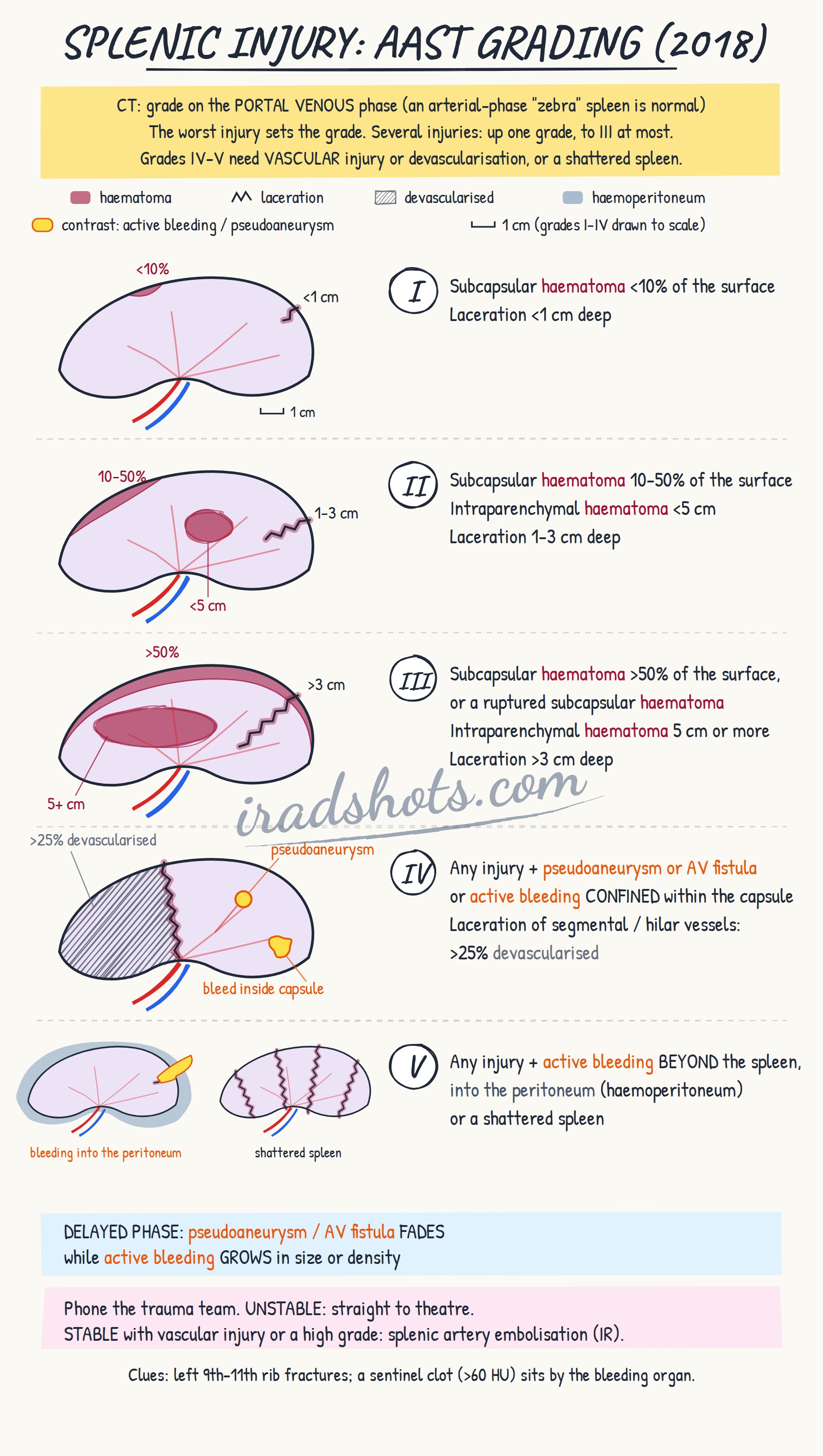
- AAST grading: Grade I (<10% subcapsular haematoma, <1 cm laceration) to Grade V (shattered spleen, or active bleeding beyond the spleen into the peritoneum)
- AAST 2018 update: vascular injury upstages severity (intrasplenic active bleed/pseudoaneurysm = Grade IV; intraperitoneal active bleed = Grade V)
- Sentinel clot sign: focal high-attenuation haematoma (>60 HU) adjacent to the spleen identifies it as the source of bleeding
- Active arterial extravasation is an irregular jet/blush that expands on delayed phases, whereas pseudoaneurysms remain well-circumscribed and follow blood pool attenuation
- Physiological 'zebra spleen' heterogeneous enhancement on early arterial phase must not be mistaken for lacerations; assess on portal venous phase
- Haemodynamically stable patients with vascular injury or high-grade tears require urgent transcatheter arterial embolisation (TAE)
The discriminators
- Physiological zebra spleen - heterogeneous enhancement seen strictly on early arterial phase that homogenises completely on portal venous phase with no perisplenic fluid.
- Splenic cleft - smooth, sharply defined cortical notch with normal adjacent parenchymal enhancement and no haematoma or hemoperitoneum.
- Splenic infarction - wedge-shaped peripheral non-enhancing defect extending to the capsule without parenchymal disruption, laceration, or sentinel clot.
Classification
AAST Splenic Injury Scale (2018 update):
Grade I: subcapsular haematoma <10% of the surface; laceration <1 cm deep.
Grade II: subcapsular haematoma 10-50% of the surface; intraparenchymal haematoma <5 cm; laceration 1-3 cm deep.
Grade III: subcapsular haematoma >50% of the surface or ruptured; intraparenchymal haematoma 5 cm or more; laceration >3 cm deep.
Grade IV: any injury with a vascular injury (pseudoaneurysm or AV fistula) or active bleeding confined within the capsule; laceration of segmental or hilar vessels with >25% devascularisation.
Grade V: any injury with active bleeding beyond the spleen into the peritoneum; shattered spleen.
Multiple injuries: advance one grade, up to grade III.
Management
Direct urgent telephone communication with the trauma team; recommend urgent transcatheter arterial embolisation (TAE) if haemodynamically stable with active extravasation, pseudoaneurysm, or high-grade injury, or immediate exploratory surgical review if unstable.
Clinical buzz words
The trap
Mistaking physiological arterial-phase heterogeneous 'zebra' enhancement for parenchymal lacerations instead of assessing the spleen on the portal venous phase.
Check yourself
1. What is the sentinel clot sign in splenic trauma and what does it indicate?
Show the answerHide the answer
2. How do you distinguish active arterial extravasation from a traumatic pseudoaneurysm in splenic injury?
Show the answerHide the answer
3. In the 2018 AAST splenic injury scale, how do vascular injuries differentiate Grade IV from Grade V?
Show the answerHide the answer
4. What is a common imaging trap when evaluating the spleen on early arterial phase CT in trauma?
Show the answerHide the answer
Cases on Radiopaedia
Find cases of splenic trauma on RadiopaediaSketch
Sign in to keep track of the lessons you have done.