Perianal fistula
Parks types, St James's grades and the anal clock; the case: a transsphincteric fistula with an ischioanal abscess.
1What the clinician needs to know
The clinician here is the colorectal surgeon. Your report must answer four questions.
- Where is the internal opening?
- How does the tract cross the sphincters? That decides whether it can be laid open safely.
- Is there hidden sepsis: a branch, an abscess, or extension above the levator? Missed sepsis brings the fistula back.
- And is it active, or a fibrotic scar?
2Anatomy and types
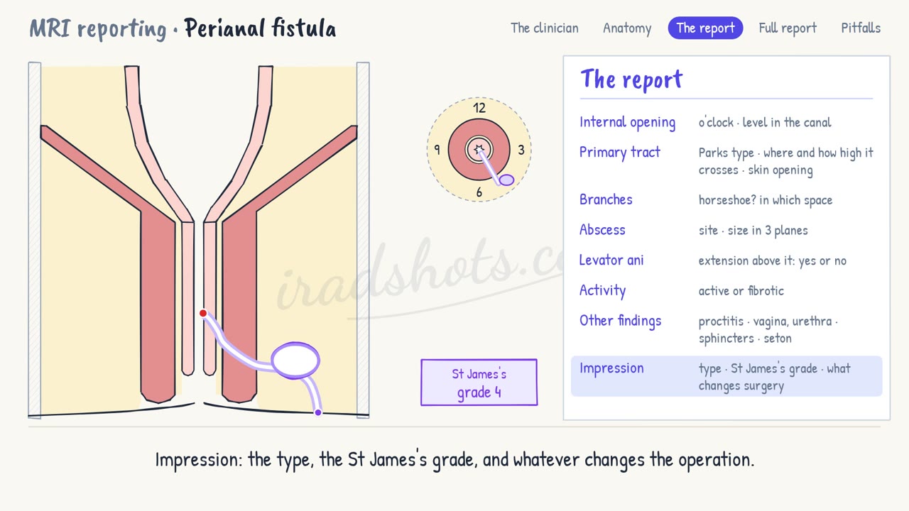
- This is the anal canal in the coronal plane, as on an oblique coronal T2 image.
- The internal sphincter is the inner ring, continuous with the circular muscle of the rectum.
- The external sphincter is the outer ring. At the top it blends with puborectalis and the levator ani.
- Between the rings lies the intersphincteric space. Outside is the ischioanal fossa, and above the levator, the supralevator space.
- The Parks types follow the main tract. Intersphincteric stays between the rings; transsphincteric crosses the external sphincter into the ischioanal fossa.
- Suprasphincteric arches over puborectalis and down through the levator. Extrasphincteric runs from the rectum, outside the sphincters altogether.
- The St James's grades build on them. Grade 1: simple intersphincteric. Grade 2: with an abscess or a branch.
- Grade 3: transsphincteric. Grade 4: with an abscess or a branch in the ischioanal fossa. Grade 5: above or through the levator.
- On axial images, 12 o'clock is anterior, 3 the patient's left, 6 posterior and 9 right.
3The report, in order
- Internal opening: its clock position and its level. Most lie at the dentate line, often posteriorly.
- Primary tract: its Parks type, where and how high it crosses the external sphincter, and its skin opening.
- Branches: any secondary tract, and any horseshoe around the canal, with its space.
- Abscess: where it is, and its size in three planes.
- Levator ani: say plainly whether there is supralevator extension. It changes the operation.
- Activity: an active tract is bright on fat-suppressed T2, with an enhancing wall. A fibrotic one is dark.
- Other findings: proctitis, a clue to Crohn's, the vagina or urethra, the sphincters, and any seton.
- Impression: the type, the St James's grade, and whatever changes the operation.
4A full report
Internal opening: 5 o'clock, at the dentate line.
Primary tract: transsphincteric, through the lower third of the external sphincter into the left ischioanal fossa. Skin opening at 4 o'clock.
Branches: none. No horseshoe.
Abscess: left ischioanal fossa, 2.4 × 1.6 × 1.5 cm.
Levator ani: intact. No supralevator extension.
Activity: active. Bright on fat-suppressed T2, with an enhancing wall.
Other findings: no proctitis. Sphincters otherwise intact.
Impression: active low transsphincteric fistula from 5 o'clock, with a left ischioanal abscess: St James's grade 4. No supralevator extension.
5Pitfalls
- Add a plain word to the clock, like 6 o'clock, posterior: surgeons examine in different positions.
- Check above the levator on the coronal images. Missed supralevator sepsis makes surgery fail.
- A tract in the ischioanal fossa is not intersphincteric. Find where it crossed.
- Dark on T2 with no enhancement is fibrosis: do not call it active.
Sign in to keep track of the reports you have done.