Breast MRI
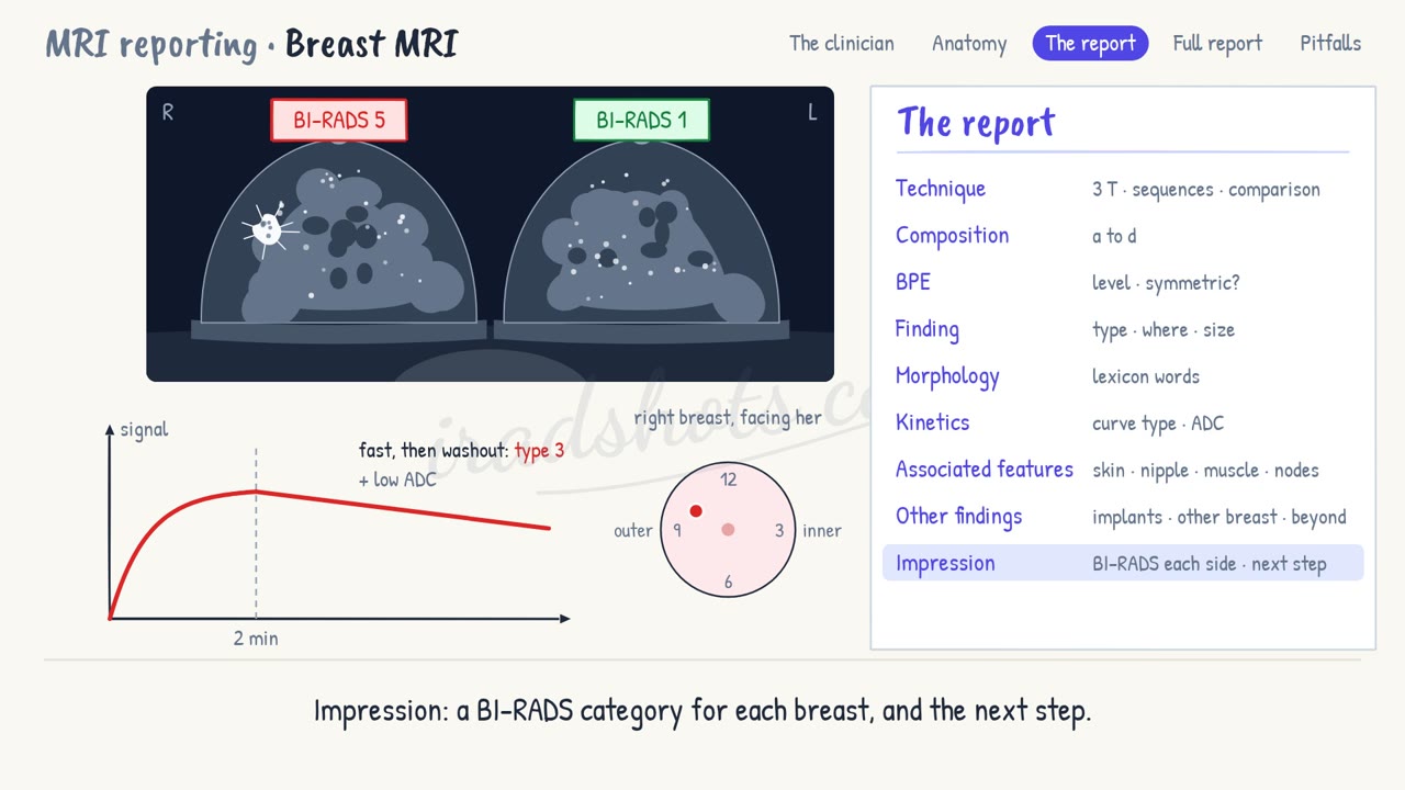
The protocol, background enhancement, mass and non-mass enhancement, the kinetic curve and BI-RADS; the case: a spiculated mass with washout.
1What the clinician needs to know
The clinician here is the breast team: the surgeon, the oncologist and the multidisciplinary meeting. Your report must answer four questions.
- Is there a suspicious lesion, and exactly where?
- How far does the disease extend, in this breast and in the other one?
- Does it reach the skin, the nipple, the pectoralis or the chest wall?
- And what is the BI-RADS category, and the next step?
2Anatomy and types
- The protocol: T1 without fat suppression shows fat; T2 shows fluid; diffusion gives the ADC; and the dynamic series is fat-suppressed T1, before and after contrast.
- Subtracting the image before contrast from the one after leaves only enhancement.
- Composition is the amount of fibroglandular tissue: a, almost entirely fat; b, scattered; c, heterogeneous; d, extreme.
- Background parenchymal enhancement, or BPE, is the normal tissue lighting up: minimal, mild, moderate or marked.
- A focus is a tiny dot, usually under 5 mm. A mass is three-dimensional, with a shape and a margin. Non-mass enhancement is neither.
- A mass has a shape: oval, round or irregular; a margin: circumscribed, irregular or spiculated; and an inside: homogeneous, heterogeneous, rim, or dark internal septations.
- Non-mass enhancement has a distribution: focal, linear, segmental, regional, multiple regions or diffuse; and a pattern: homogeneous, heterogeneous, clumped or clustered ring.
- For the curve, put a small region of interest on the brightest part of the lesion, and plot its signal over time.
- In the first 2 minutes, the initial phase rises slowly, at a medium rate, or fast.
- The delayed phase gives the type. Type 1 keeps rising: persistent, usually benign. Type 2 flattens: plateau, indeterminate. Type 3 falls: washout, suspicious.
- The BI-RADS categories: 1 negative, 2 benign, 3 probably benign, 4 suspicious, 5 highly suggestive of cancer, 6 known cancer; and 0 when more imaging is needed.
3The report, in order
- Technique: field strength, sequences, contrast, and any comparison.
- Composition: the amount of fibroglandular tissue, a to d.
- BPE: its level, and whether it is symmetric.
- Finding: focus, mass or non-mass; side, clock face, depth and distance from the nipple; and size in three planes.
- Morphology: the descriptors, in the words of the lexicon.
- Kinetics: the initial rise and the delayed curve; and the ADC, if diffusion was done.
- Associated features: skin, nipple, pectoralis, chest wall and lymph nodes.
- Other findings: non-enhancing lesions, implants, the other breast, and anything outside the breasts.
- Impression: a BI-RADS category for each breast, and the next step.
4A full report
Technique: 3 T, dynamic contrast-enhanced with subtraction, and diffusion. No prior MRI.
Composition: c, heterogeneous fibroglandular tissue.
BPE: mild and symmetric.
Finding: right breast, mass at 10 o'clock, middle depth, 6 cm from the nipple, 14 × 11 × 12 mm.
Morphology: irregular shape, spiculated margin, heterogeneous internal enhancement.
Kinetics: fast initial rise, then washout: type 3. Restricted diffusion.
Associated features: none. Skin, nipple, pectoralis and axillary nodes normal.
Other findings: no implants. The left breast and the rest of the field are unremarkable.
Impression: right breast BI-RADS 5, an irregular spiculated mass with washout at 10 o'clock. Left breast BI-RADS 1. Second-look ultrasound and biopsy.
5Pitfalls
- Morphology outranks kinetics: a suspicious shape needs biopsy, even with a persistent curve.
- Put the region of interest on the most enhancing part, not on the whole lesion or a vessel.
- Lymph nodes wash out too: look for the fatty hilum before calling a node suspicious.
- Marked BPE can hide or mimic disease: scan premenopausal women in the second week of the cycle.
Sign in to keep track of the reports you have done.