Degenerative lumbar spine
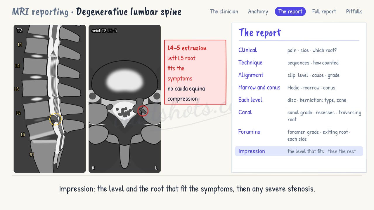
Disc degeneration, bulges and herniations, their zones and the roots they press on, canal and foraminal stenosis, Modic changes and slips; the case: an L4-5 extrusion.
1What the clinician needs to know
The clinician here is the spinal surgeon, or the pain team. Your report must answer four questions.
- Which level, and which nerve root, explain the pain?
- Is it a disc, a narrow canal, or a narrow foramen?
- How severe is the narrowing, and is the cauda equina compressed?
- Is there a slip, which may change the operation?
2Anatomy and types
- A disc is a soft nucleus inside a tough ring, the annulus. With age it dries out: on T2 it darkens, and it loses height.
- Pfirrmann grades this from 1, a bright disc of normal height, to 5, a dark and collapsed one.
- A bulge is disc tissue spread beyond the edges of the vertebra, over more than a quarter of the disc's circumference. It is common with age, and it is not a herniation.
- A herniation is focal: under a quarter of the circumference. In a protrusion, its base is wider than what sticks out.
- In an extrusion, what sticks out is wider than its base, or it runs up or down beyond the disc. A sequestration has lost contact with the disc altogether.
- Name the zone it reaches: central, subarticular in the lateral recess, foraminal, or extraforaminal.
- At L4-5, a subarticular herniation presses on the traversing root, L5. A foraminal or extraforaminal one presses on the exiting root, L4.
- The canal narrows from the disc in front, and from thick ligamentum flavum and facet joints behind. On axial T2, grade it by the CSF left around the roots.
- Mild still has CSF between the roots. Moderate has the roots crowded together, with a little CSF. Severe has no CSF left at all.
- In the foramen, the root sits in fat. Fat lost on two opposite sides is mild; fat lost all round is moderate; a squashed root is severe.
- At the endplates, Modic changes: type 1 is oedema, dark on T1 and bright on T2; type 2 is fat, bright on both; type 3 is sclerosis, dark on both.
- Worn facet joints can let a vertebra slip forward: a degenerative spondylolisthesis, usually at L4-5. Meyerding grades the slip by quarters, from 1 to 4.
3The report, in order
- Clinical: the pain, its side, and the root it seems to come from.
- Technique: the sequences, and how the levels were counted.
- Alignment: any slip, with its level, its cause and its grade.
- Marrow and conus: Modic changes, the marrow, and where the conus ends.
- Each level: the disc, and any herniation, with its type and zone.
- Canal: At that level, the canal and the lateral recesses: the grade, and any traversing root that is pressed on.
- Foramina: its grade, and the exiting root.
- Impression: the level and the root that fit the symptoms, then any severe stenosis.
4A full report
Clinical: Left leg pain for eight weeks, in an L5 distribution. No red flags.
Technique: Sagittal T1, T2 and STIR, and axial T2 from L3 to S1. Five lumbar vertebrae, counted down from C2.
Alignment: Normal.
Marrow and conus: Modic type 2 change at L5-S1. Otherwise normal marrow. The conus ends at L1.
L1-2 and L2-3: Normal.
L3-4: Disc bulge. No stenosis.
L4-5: Left subarticular disc extrusion, migrating 6 mm down. Moderate canal stenosis. Severe left lateral recess stenosis, compressing the traversing left L5 root. Patent foramina.
L5-S1: Disc degeneration, Pfirrmann 4, with a bulge. Mild narrowing of both foramina; the exiting L5 roots are not compressed.
Impression: Left subarticular disc extrusion at L4-5, compressing the traversing left L5 root, which fits the symptoms. No cauda equina compression.
5Pitfalls
- Do not call every disc a bulge: a focal herniation that presses on a root must be named as one.
- Name the root and its path: a traversing L5 root and an exiting L4 root are reached by different approaches.
- Look above and below the disc for a migrated or sequestered fragment: the surgeon must know where it is.
- Modic type 1 can look like discitis. Discitis has a bright disc, eroded endplates and a collection.
Sign in to keep track of the reports you have done.