Lumbar spine
Counting the levels, the sequences, the marrow on T1, the conus and cauda equina, the red flags and what lies beyond the spine; the case: a lesion in the marrow.
1What the clinician needs to know
The clinician here may be a GP, a spinal surgeon, or the emergency team. Your report must answer four questions.
- Is there a red flag: cauda equina compression, infection, a tumour or a fracture?
- Is there a cause that fits the symptoms, such as a nerve root that matches the leg pain?
- At exactly which level, counted so that the surgeon finds the same one?
- And does the cord end normally, and is there anything beyond the spine?
2Anatomy and types
- Five lumbar vertebrae sit on the sacrum. A level is named by its disc, such as L4-5, and each nerve root leaves under the pedicle of its own vertebra.
- Count the vertebrae from a fixed point: down from C2 on the localiser, or up from the sacrum. Then say in the report how you counted.
- A transitional vertebra is common: a sacralised L5, or a lumbarised S1. Describe it, because it changes the numbering.
- The sequences are sagittal T1, T2 and STIR, and axial T2 through the lower discs. T1 shows the marrow, T2 the discs and the nerves, and STIR shows oedema.
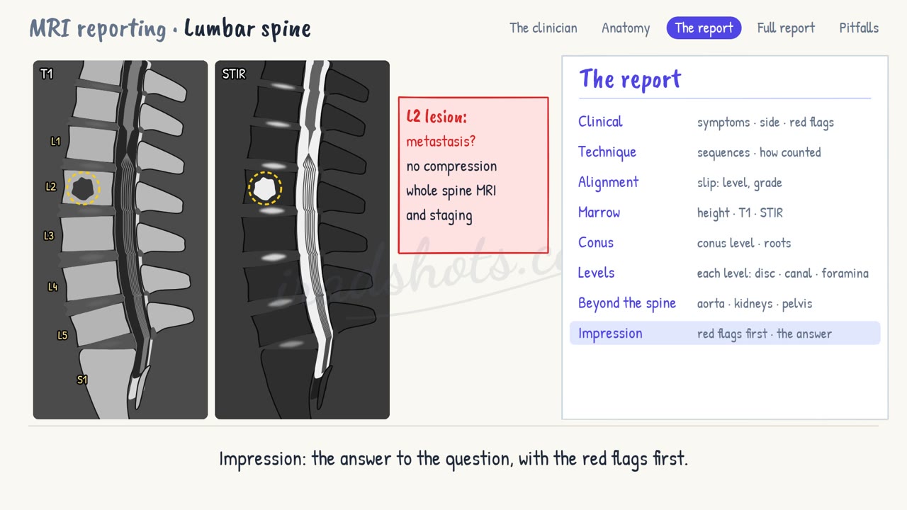
- On T1, normal adult marrow contains fat, so it is brighter than the discs. A vertebra darker than its disc on T1 has lost its fat: to tumour, infection or a fracture.
- The cord ends at the conus, normally at L1 or L2. A conus below the L2-3 disc is low lying: think of a tethered cord.
- Below it, the nerve roots of the cauda equina float in CSF. Roots crowded with no CSF around them, at a narrow level, are cauda equina compression: phone it through.
- Infection, discitis, makes the disc bright on T2 and STIR, erodes both endplates, and may form a collection in front of the spine or in the epidural space.
- A metastasis replaces the marrow: dark on T1, and often reaching the pedicle. An epidural mass may press on the cord or the cauda equina.
- An osteoporotic fracture keeps some fatty marrow, with a fracture line. A pathological fracture replaces the whole body, and bulges its back wall.
- Last, look beyond the spine on every scan: the aorta, the kidneys and the pelvis.
3The report, in order
- Clinical: the symptoms, their side, and any red flags.
- Technique: the sequences, and how the levels were counted.
- Alignment: normal, or the level and grade of any slip.
- Marrow: the height of the vertebrae, and the marrow signal on T1 and STIR.
- Conus: where it ends, and the nerve roots of the cauda equina.
- Levels: from L1-2 down to L5-S1, the disc, the canal and the foramina.
- Beyond the spine: the aorta, the kidneys and the pelvis.
- Impression: the answer to the question, with the red flags first.
4A full report
Clinical: Low back pain for six weeks, worse at night. Previous breast cancer.
Technique: Sagittal T1, T2 and STIR, and axial T2. Five lumbar vertebrae, counted down from C2.
Alignment: Normal.
Marrow: A 2 cm lesion in the L2 vertebral body, dark on T1 and bright on STIR, reaching the right pedicle. No collapse. Normal marrow elsewhere.
Conus: Ends at L1. Normal cauda equina.
Levels: Mild disc degeneration at L4-5 and L5-S1. No canal or foraminal stenosis.
Beyond the spine: Normal aorta and kidneys.
Impression: Lesion in the L2 vertebral body and right pedicle, suspicious for a metastasis. No epidural extension and no neural compression. Suggest MRI of the whole spine and staging, with the oncology team.
5Pitfalls
- Count from a fixed point and say how: a wrong level means surgery at the wrong level.
- Read the marrow on T1 before the discs: a metastasis is easy to see first, and easy to miss after.
- Follow the cord to the conus on every scan: a low conus, or a tumour of the conus, hides at the top.
- Look beyond the spine: an aortic aneurysm or a kidney tumour can be the cause of back pain.
Sign in to keep track of the reports you have done.