mpMRI prostate
Zones and sectors, PSA density, PI-RADS v2.1 in the peripheral and transition zones, and staging; the case: a PI-RADS 5 lesion.
1What the clinician needs to know
The clinician here is the urologist, and the prostate cancer team. Your report must answer four questions.
- Is there a lesion that is likely to be significant cancer?
- Where exactly is it, so that a biopsy can target it?
- Has it spread beyond the prostate, into the fat or the seminal vesicles?
- And are there suspicious nodes, or bone metastases?
2Anatomy and types
- The prostate has four zones: the peripheral zone, at the back and sides, where most cancers start; the transition zone, around the urethra, where BPH grows; the central zone, at the base; and the anterior fibromuscular stroma, in front.
- From top to bottom, the gland has a base, a mid-gland and an apex. PI-RADS splits each level into sectors, so every lesion has an address.
- Measure the gland in three directions and multiply by 0.52, for its volume. The PSA divided by the volume is the PSA density: above 0.15 is a warning sign.
- On T2, the normal peripheral zone is bright, and cancer is dark. On diffusion, cancer is dark on the ADC map, and bright on the high b value image, of at least 1400.
- PI-RADS scores each lesion from 1 to 5: how likely it is to be clinically significant cancer, meaning Gleason 3 + 4 or more.
- In the peripheral zone, diffusion decides. Normal scores 1; a linear or wedge-shaped area, 2; a focal area, but not marked on both maps, 3.
- Focal and marked on both, dark on ADC and bright on high b value, scores 4; and 5 if it is 1.5 cm or more, or goes beyond the gland.
- Dynamic contrast only breaks a tie: in the peripheral zone, a 3 that enhances early and focally becomes a 4.
- In the transition zone, T2 decides. A round, fully encapsulated nodule scores 1; a mostly encapsulated one, 2; a heterogeneous area with blurred margins, 3.
- A lens-shaped or ill-defined, homogeneous, moderately dark area scores 4; and 5 if it is 1.5 cm or more, or goes beyond the gland.
- Here, diffusion can lift the score: a 2 becomes a 3 if diffusion scores 4 or 5, and a 3 becomes a 4 if diffusion scores 5.
- Then the edge of the gland. Extraprostatic extension: a bulge or a break in the capsule, contact with it over 1 cm, an asymmetric neurovascular bundle, or an obliterated rectoprostatic angle.
- Seminal vesicle invasion: dark T2 signal, restricted diffusion or enhancement in a vesicle, or tumour growing into it from the base.
- Last, the pelvic nodes: over 8 mm in short axis is suspicious; and the bones, for metastases.
3The report, in order
- Clinical: the PSA, and any previous biopsy, with its date.
- Technique: the scanner, the sequences with the highest b value, and the image quality.
- Gland: its three measurements, the volume, and the PSA density.
- Lesion: its address, by zone, side, level and sector; and its size, on ADC in the peripheral zone, on T2 in the transition zone.
- Score: what T2, diffusion and contrast show, and the PI-RADS score. In the UK, NICE also asks for a Likert score.
- Staging: extraprostatic extension and its side, the neurovascular bundles, and the seminal vesicles.
- Nodes and bones: the pelvic nodes, the bones, and the rest of the pelvis.
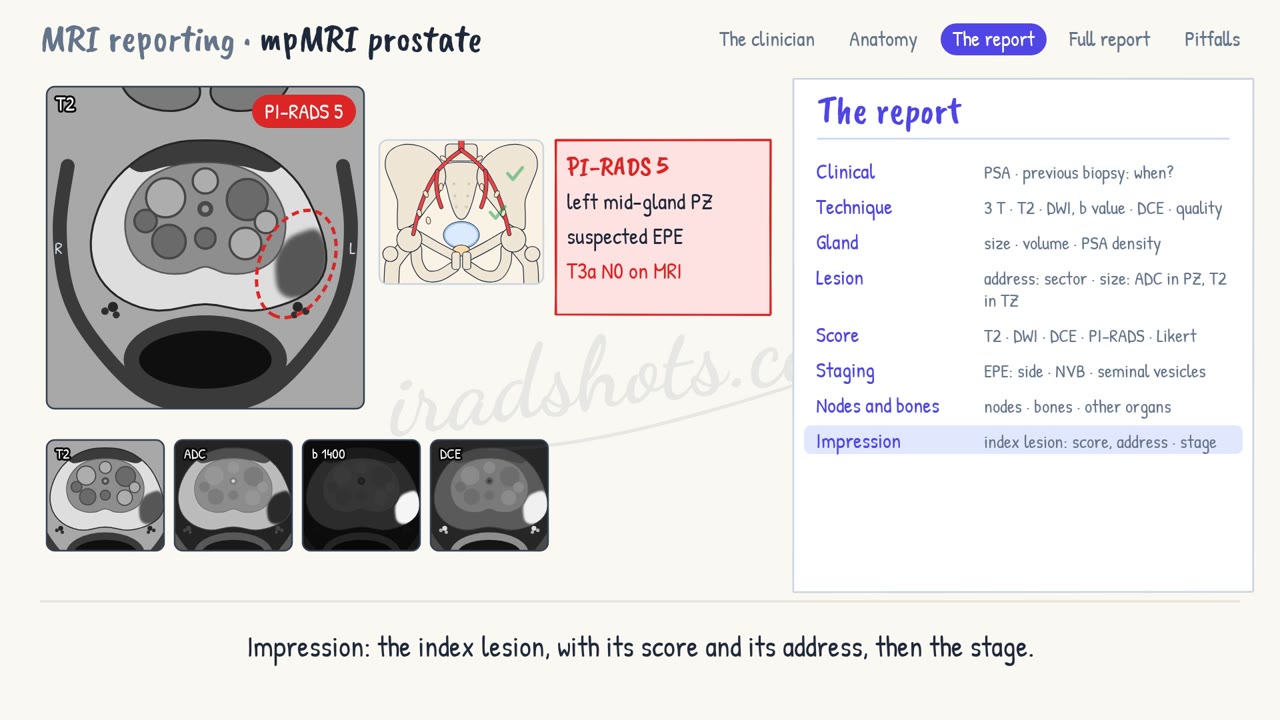
- Impression: the index lesion, with its score and its address, then the stage.
4A full report
Clinical: PSA 8 ng/mL. No previous biopsy.
Technique: 3 T. T2, diffusion with b 1400, and dynamic contrast. Good image quality.
Gland: 5.0 × 3.7 × 4.2 cm, volume 40 mL. PSA density 0.20.
Lesion: left mid-gland, peripheral zone, posterolateral; 17 mm on ADC.
Score: dark on T2, markedly restricted on diffusion, with early focal enhancement. PI-RADS 5; Likert 5.
Staging: the lesion bulges the capsule, with 14 mm of contact: suspected left extraprostatic extension. Neurovascular bundles spared. Seminal vesicles clear.
Nodes and bones: no suspicious pelvic nodes. No bone metastases.
Impression: PI-RADS 5 lesion in the left mid-gland peripheral zone, with suspected extraprostatic extension: T3a N0 on MRI. Suitable for targeted biopsy.
5Pitfalls
- After a biopsy, blood is dark on T2 and can mimic tumour: look for it on T1, where it is bright.
- The central zone is dark on T2 and ADC, at the base, on both sides: symmetry says it is normal.
- Look hard at the front of the gland: anterior tumours hide among BPH nodules.
- Give every lesion a full address: a lesion the urologist cannot find cannot be biopsied.
Sign in to keep track of the reports you have done.