MR defecography
Evacuation, the PCL, the H and M lines, the anorectal angle and each compartment; the case: an anterior rectocele that holds gel.
1What the clinician needs to know
The clinician here is the pelvic floor team: colorectal surgeons, urogynaecologists and physiotherapists. Your report must answer four questions.
- Does the rectum empty, and how much is left?
- Which compartments drop, and how far?
- Is there a rectocele, an intussusception or an enterocele?
- And is the floor weak, or is the muscle failing to relax?
2Anatomy and types
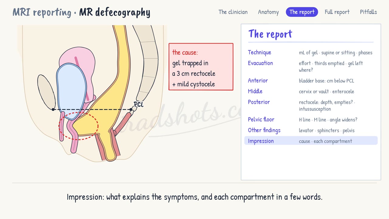
- This is the female pelvis in the midline, in three compartments: anterior, with the bladder and urethra; middle, with the vagina and uterus; and posterior, with the rectum.
- The rectum is filled with gel. Cine images are taken at rest, on squeezing, and above all during evacuation, repeated until the rectum is empty.
- The reference is the pubococcygeal line, or PCL: from the lower edge of the symphysis to the last joint of the coccyx, not its tip.
- Each organ is measured at right angles to it: the bladder base, the cervix or vault, and the anorectal junction. The rule of three grades the drop: under 3 cm mild, 3 to 6 moderate, over 6 severe.
- The H line runs from the symphysis to the anorectal junction: the width of the hiatus. Normal on straining is up to 6 cm.
- The M line drops from the PCL to the end of the H line: how far the floor descends. Normal is up to 2 cm.
- The anorectal angle, between the anal canal and the back wall of the rectum, narrows on squeezing, and widens on evacuation.
- A rectocele is the front wall of the rectum bulging forward. Measure how far it bulges beyond the line of the anal canal, and say whether it empties.
- Intussusception is the rectal wall folding in: intrarectal, intra-anal, or external, which is a rectal prolapse.
- An enterocele is small bowel dropping between the vagina and the rectum, often only at the end of evacuation.
- In pelvic floor dyssynergia, the puborectalis tightens instead of relaxing: the angle narrows, and the rectum does not empty.
3The report, in order
- Technique: the volume of gel, the position, and the phases.
- Evacuation: the patient's effort, how much emptied, in thirds, and where gel is left.
- Anterior: the bladder base, in centimetres below the PCL.
- Middle: the cervix or vault, and any enterocele.
- Posterior: a rectocele, its depth and whether it empties; and any intussusception.
- Pelvic floor: the H and M lines, and whether the anorectal angle widens or narrows.
- Other findings: levator tears, the sphincters, and anything else in the pelvis.
- Impression: what explains the symptoms, and each compartment in a few words.
4A full report
Technique: supine, 120 mL of rectal gel. Sagittal cine at rest, on squeezing and during three evacuations.
Evacuation: good effort. Two thirds emptied; the rest is held in the rectocele.
Anterior compartment: bladder base 1 cm below the PCL: mild cystocele.
Middle compartment: cervix 1 cm above the PCL. No enterocele.
Posterior compartment: anterior rectocele, 3 cm deep, holding gel. No intussusception.
Pelvic floor: H line 7 cm and M line 2.5 cm: a widened hiatus and descent. The anorectal angle widens normally, from 105° to 130°.
Other findings: levator ani and anal sphincters intact.
Impression: incomplete evacuation, with gel trapped in an anterior rectocele, 3 cm deep. Mild cystocele and pelvic floor descent. No intussusception, enterocele or dyssynergia.
5Pitfalls
- Never stop at straining: without evacuation, prolapse is underestimated and enteroceles are missed.
- Say how hard the patient tried: poor effort hides prolapse, and can look like dyssynergia.
- Draw the PCL to the last coccygeal joint, not to the tip of the coccyx.
- Report all three compartments: prolapse is often in more than one, and repairing one can unmask another.
Sign in to keep track of the reports you have done.